Affordable, high-resolution, medical diagnostics imaging using dielectric spectroscopy
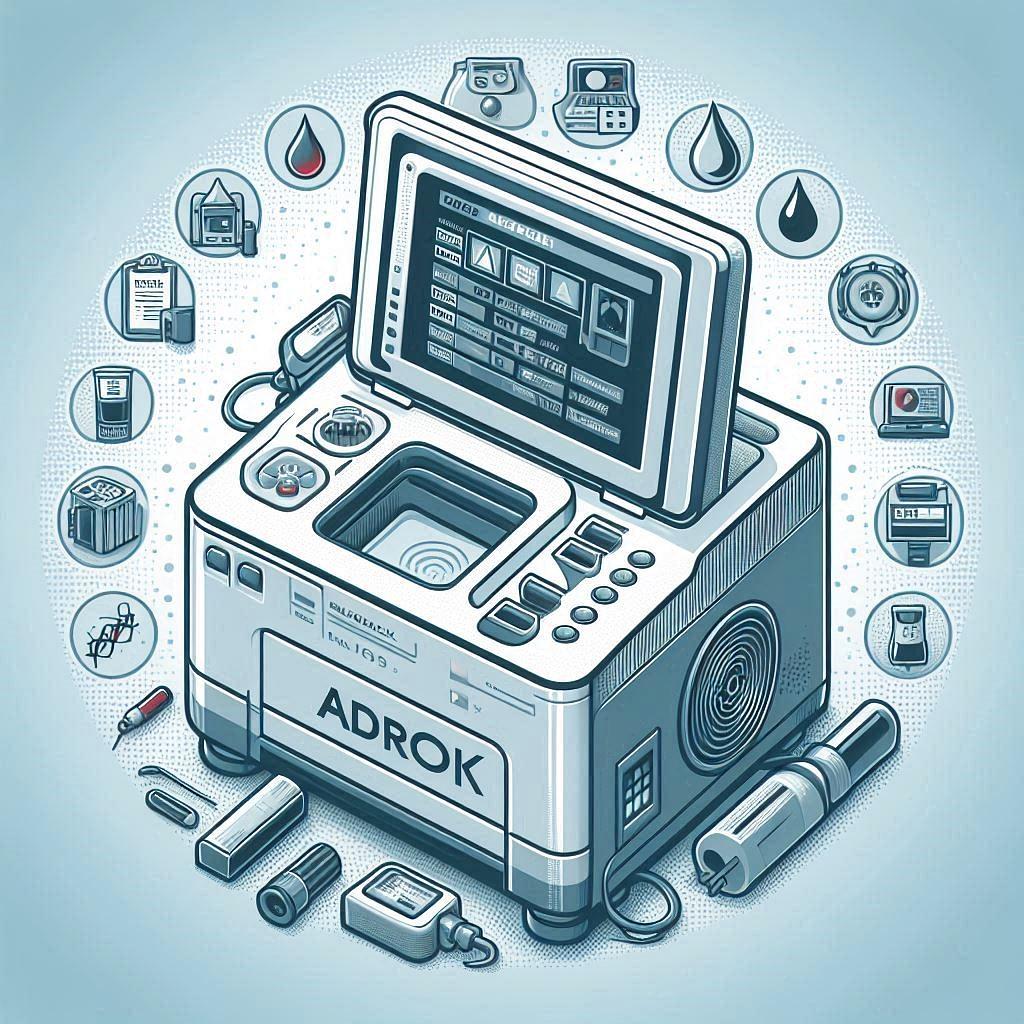
Although ADR is primarily geological, there has been experimental exploration of its potential in medical applications. For instance:
- A laboratory-based study used ADR to analyze blood samples from patients with Creutzfeldt–Jakob disease (CJD), a prion disease. The technique aimed to distinguish infected samples from controls by their dielectric response.
Applying ADR to Blood Diagnostics
When ADR spectroscopy is applied to blood samples, the technology is measuring:
- Dielectric permittivity (ability to store electric energy)
- Conductivity (ability to carry current)
- Resonant frequencies (unique peaks tied to molecular structure and composition)
Changes in blood composition caused by disease (e.g., altered proteins, abnormal cells, or pathogen presence) shift these dielectric signatures.
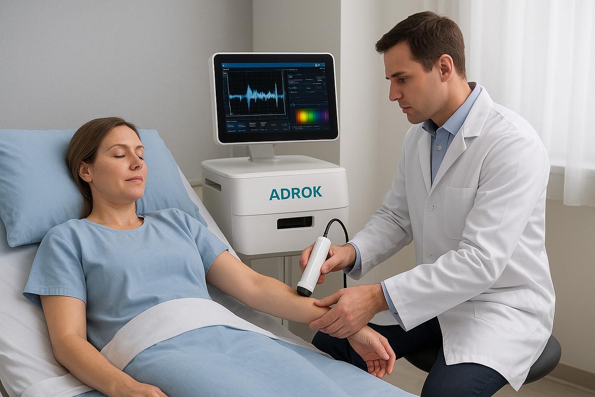
Research Example: Creutzfeldt–Jakob Disease (CJD)
- ADR spectroscopy has been tested on blood samples from patients with CJD (a prion disease).
- In those studies, ADR could distinguish between infected and healthy samples by detecting subtle shifts in dielectric resonance signatures.
- This suggested the potential for ADR as a non-invasive, rapid screening tool for otherwise hard-to-diagnose conditions.
Why Blood Diagnostics?
Blood is ideal for ADR spectroscopy because:
- It contains proteins, electrolytes, and cells — each with unique dielectric properties.
- Diseases often alter blood chemistry (e.g., abnormal proteins in cancer, misfolded proteins in neurodegenerative diseases, or structural changes in red blood cells in sickle cell disease).
- ADR could, in principle, detect these changes without chemical reagents or staining — just by analyzing the electromagnetic response.
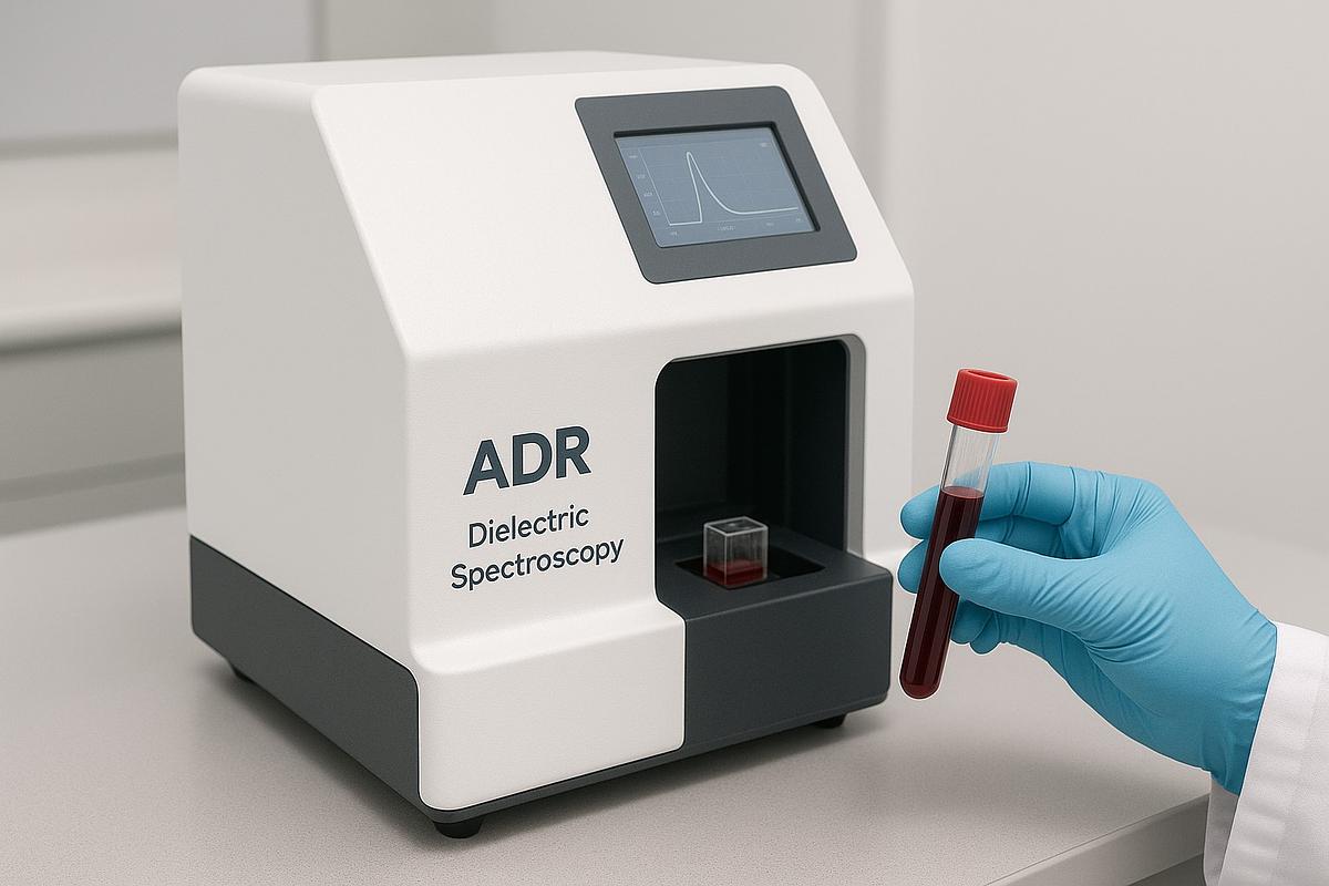
Advantages & Limitations
✅ Advantages:
- Non-invasive and label-free (no dyes, no radioactive tracers).
- Potential for early detection of disease-related changes.
- Fast measurements.
⚠️ Limitations:
- Still experimental — not yet standardized for clinical use.
- Requires careful calibration and large-scale validation.
- Disease signals may overlap with normal biological variability.
ADR spectroscopy for blood diagnostics is about detecting disease-specific changes in the electromagnetic “fingerprint” of blood. It has shown promise in pilot studies (like CJD), and conceptually could extend to cancers, hematologic disorders, or infections, but it’s still an emerging, exploratory method rather than a routine medical test.
- ADR’s strength = broad, label-free screening — it detects abnormal dielectric signatures without needing a specific antibody or gene sequence. This makes it potentially useful for diseases with unknown or complex biomarkers (e.g., prion diseases, early cancer states).
- Traditional methods = high specificity — ELISA, PCR, and flow cytometry excel at pinpointing known molecules or cells, but require prior knowledge of the target and lab infrastructure.
- Best fit for ADR = a frontline screening tool: fast, portable, and able to flag “something is wrong,” which can then be confirmed with conventional, highly specific methods.
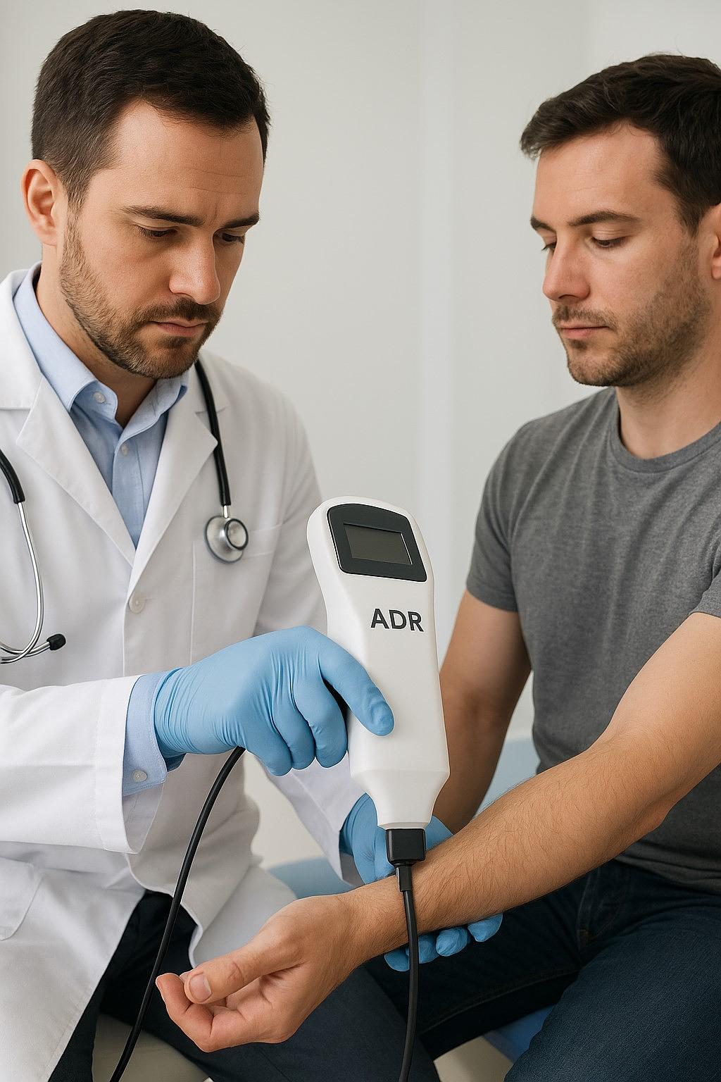
Dielectric Spectroscopy in Medical Diagnostics
What is dielectric spectroscopy? Dielectric spectroscopy, also referred to as electrical impedance spectroscopy or bioimpedance when applied to tissue or cells, is a technique that applies varying-frequency electrical signals to biological materials and measures how they respond—yielding information about properties like permittivity, conductivity, and cell membrane capacitance. It's non-invasive, label-free, and can often yield results in real-time.
How is it being used medically?
- Cancer diagnosis: Multiple studies have found that dielectric properties differ noticeably between normal and cancerous tissues—across skin, oral cavity, cervical, bladder, breast, liver, thyroid, lung, and prostate tumors - likely due to differences in composition, blood flow, and tissue architecture. These dielectric parameters have been proposed as potential biomarkers for early detection and surgical guidance
- Sickle Cell Disease: Advanced impedance spectroscopy in microfluidic systems has shown that red blood cells impacted by hypoxia (low oxygen) exhibit measurable changes - such as higher interior permittivity and conductivity and reduced membrane capacitance - allowing discrimination between sickle and normal cells
- Cell/tissue stress & cancer: Broadband dielectric spectroscopy has been used to monitor structural and biophysical changes in tissues - such as from radiation exposure or developing cancers (e.g. lung, rostate) - highlighting unique dielectric signatures tied to damage or malignancy.
- Corneal assessments: Dielectric spectroscopy has been used in porcine corneal studies to understand effects of hydration and temperature, offering a non-invasive way to detect structural variations.
- Biofluid analyses: CMOS-based dielectric biosensors have detected differences in dielectric properties of biofluids (e.g. blood, saliva, semen) between diseased and healthy subjects - useful in point-of-care diagnostics.
- General real-time cell sensing: Platforms like electrical cell substrate impedance sensing (ECIS) are employed to monitor cell behaviors (like differentiation, division, cytotoxicity) in real-time.
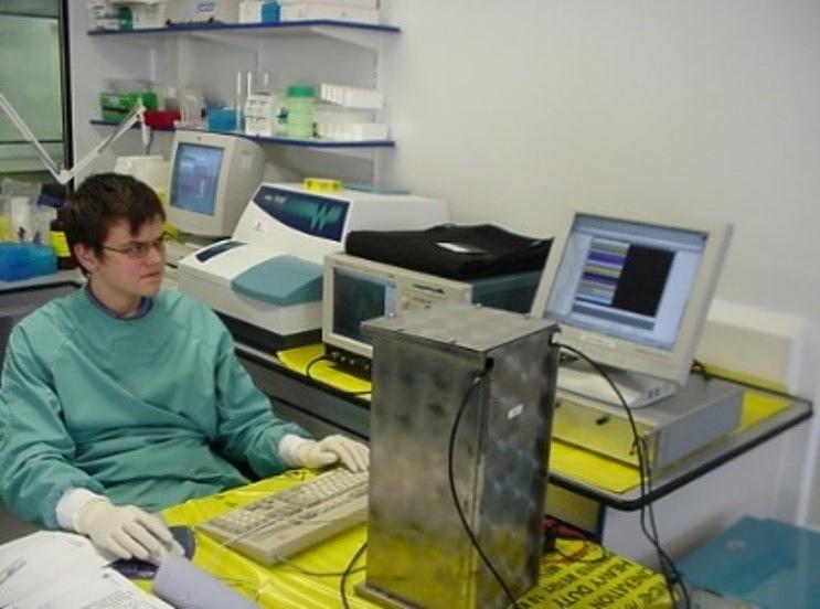
Adrok’s ADR: Primarily a geophysical tool, ADR has shown early-stage promise in differentiating diseased from non-diseased biological samples, such as in prion diseases (CJD). Its specialty lies in detecting dielectric signatures in a lab setting.
Dielectric Spectroscopy (EIS/Bioimpedance): A growing and versatile diagnostic method already applied across numerous medical fields - from cancer and hematology to ophthalmology - by non-invasively detecting changes in dielectric properties at cellular and tissue levels.
Dielectric spectroscopy (EIS/bioimpedance) has far broader real-world application and validation in medical diagnostics so far. Adrok’s ADR represents an innovative, exploratory application with potential, but it remains more experimental - showing specific interest in re-purposing geophysical techniques for biological analysis.
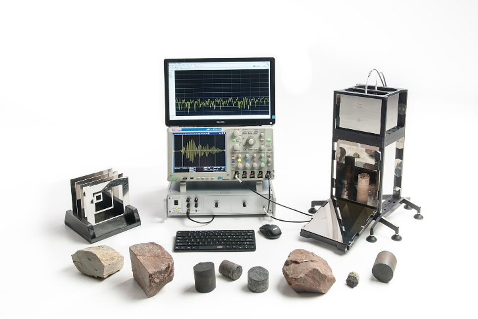
The technology is based on assessing the transmission, reflection and absorption image properties of objects in ultra-wide bands at invisible light frequencies (radio-wave, micro-wave, sub-micro-wave and thermal infra-red wave-bands). These waves, when passing through materials, cause the electrons within the atoms or molecules to resonate. The resonance exhibited by any given material has been found to differ from all other materials. By typecasting and building a library of such resonance responses, ADROK aim progressively to detect a wide variety of materials.
In addition to its capability to detect materials and image with a high degree of precision, ADR technology has other inherent advantages. The energy employed is very low, and the waves generated are non-destructive, so there is no detectible chemical or biological change in the materials under investigation. The waves can operate at microscopic ranges to pick out changes at the micron level. The equipment can be used from a static or moving platform either on the surface of land or water or, potentially, from airborne platforms. The equipment itself is light, compact and readily transportable.
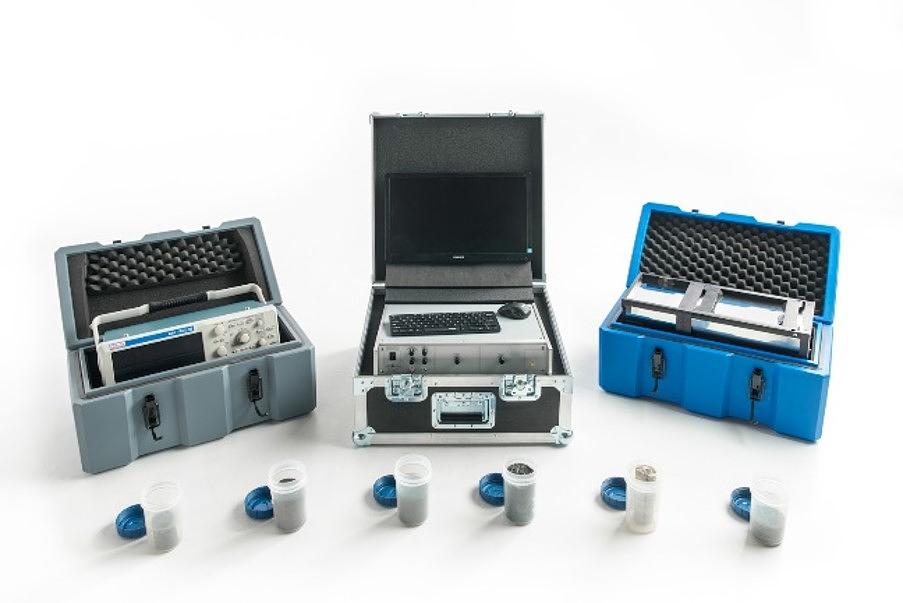
ADR technology has the potential to displace or complement established 2-D and 3-D diagnostic imaging modalities, such as x-ray, magnetic resonance imaging, CT and ultrasound. It has also demonstrated the ability to non-invasively identify and quantify biological material both in-vitro and in-vivo. The ultimate extent to which ADR will impact on existing imaging and diagnostic methods is not yet known but is predicted to be high. In common with all revolutionary technologies, developments and refinements to extend its capability will continue for decades.
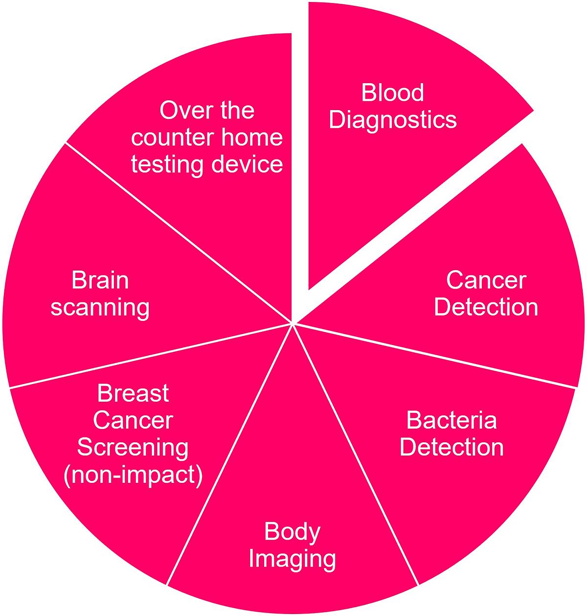
Initial plot studies in life sciences diagnostics have shown great promise using non-ionising, non-destructive ADR prototype systems which detected some alteration in physiology reflecting in some characteristic change in overall blood composition (probably too subtle, and probably complex multicomponent, to be picked up by measuring only one component by conventional tests). Adrok propose that there will be characteristic blood chemistries unique to different diseases which your technology may be capable of recognising.
Adrok are seeking investors and partners to help develop commercial Adrok Health technology. Get in touch if you would like to help.
Selected Adrok Publications
1.Fagge, T.J., Barclay, G.R., Stove, G.C., Stove, G., Robinson, M.J., Head, M.W., Ironside, J.W. and Turner, M.L., Application of Atomic Dielectric Resonance Spectroscopy for the screening of blood samples from patients with clinical variant and sporadic CJD. J. Translational Medicine, 2007, 5:41. DOI: 10.1186/1479-5876-5-41
2.Valentine, H., Fraser, J., Stove, G.C., Barclay, G.R. and Farquhar, C.F. Atomic dielectric resonance spectroscopy: a novel technique for the identification of TSE infected tissues. International Conference on Transmissible Spongiform Encephalopathies, Edinburgh, 2002/9
3.van den Doel, K. and Robinson, M., Numerical Simulation of Aquifer Detection Using Low Frequency Pulsed Radar, PIERS 2015, Prague.
4.van den Doel, K., Jansen, J., Robinson, M., Stove, G.C. and Stove, G.D.C.,Ground penetrating abilities of broadband pulsed radar in the 1-70MHz range. In: SEG Technical Program Expanded Abstracts 2014, Denver. 1770–1774.
5.van den Doel, K. and Stove, G., Modeling and Simulation of Low Frequency Subsurface Radar Imaging in Permafrost. Computer Science and Information Technology, 2018 6(3), 40–45.

